
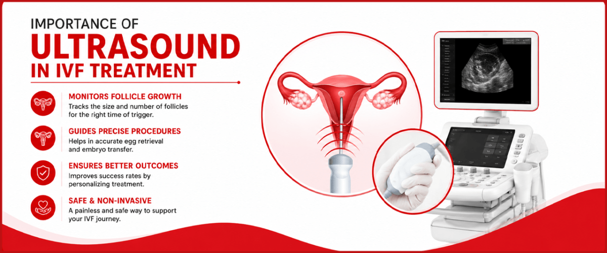
The Importance of Ultrasound in IVF: Imprimis IVF
Importance of Ultrasound in IVF: So much of IVF happens where no one can see it: eggs ripening deep inside the ovaries, a lining quietly thickening, an embryo settling into the womb. It can feel like you’re trusting a process you can’t watch. Except your doctor can watch it, in real time, and the tool that lets them is one you’ll come to know very well: ultrasound.
If IVF has a pair of eyes, this is it. No single technology is used more often across a cycle, and few decisions are made without it. Every dose adjustment, the exact day of your trigger shot, the safe collection of your eggs, the moment your embryo is placed- ultrasound guides them all. Understanding what those scans are doing turns you from a passenger into a partner in your own treatment.
Here’s how ultrasound works at every stage, what the numbers mean, and why it matters so much for your chances.
Why Ultrasound is the Backbone of IVF
Ultrasound uses harmless sound waves, not radiation, to build a live picture of your ovaries, follicles, and uterus. It’s fast, it’s radiation-free, it can be repeated as often as needed, and it shows changes as they happen, which is exactly what a fast-moving IVF cycle demands.
You’ll meet it in two forms. A transvaginal ultrasound, where a slim probe gives a close, detailed view of the ovaries and lining, is the everyday workhorse of IVF. A transabdominal ultrasound, done over the belly (usually with a full bladder), is often used to guide the embryo transfer. Between them, they cover everything from your first assessment to your first glimpse of a heartbeat.
Understanding the Importance of Ultrasound in IVF helps patients see how ultrasound supports monitoring and decision-making throughout the treatment cycle.
Importance of Ultrasound in IVF at Every Stage
The Importance of Ultrasound in IVF becomes clear when you follow its role from the baseline scan through stimulation, egg retrieval, embryo transfer, and early pregnancy monitoring.
Before you start, the baseline scan
Before any medication, a baseline transvaginal scan takes stock. The key measurement is your antral follicle count, the number of small resting follicles visible on the ovaries, which, alongside your AMH blood level, estimates your ovarian reserve and helps your specialist choose the right medication dose. The same scan checks your uterus for fibroids, polyps, or shape abnormalities, and your ovaries for cysts or the many-small-follicle pattern typical of PCOS. It can also flag a fluid-filled tube (hydrosalpinx) that could lower success. In short, this one scan personalises your entire protocol and catches problems that could quietly block implantation later.
During stimulation follicle tracking
Once your stimulation injections begin, ultrasound becomes a regular companion, usually a scan every two to three days, often three to six across the cycle. Each one measures how many follicles are growing and how big they are, so your doctor can fine-tune your medication in real time: too little response, and the dose goes up; too much, and they act to protect you from ovarian hyperstimulation. The lining is watched at the same time. This constant feedback loop is why no two IVF cycles are dosed exactly alike.
Timing the trigger
The whole cycle builds to one precisely timed moment: the trigger shot that matures your eggs. Ultrasound is what determines it. When the leading follicles reach roughly 18 to 20 mm, they’re judged mature, and the trigger is scheduled. Call it a day too early and the eggs aren’t ready; too late and you may lose them to ovulation. That single measurement, made on screen, sets the clock for everything that follows.
Egg retrieval: the live guide
During egg retrieval, ultrasound isn’t just monitoring; it’s steering. A transvaginal probe with a fine needle guide lets your specialist see each follicle on screen and pass the needle through the vaginal wall directly into it, one by one, drawing out the fluid that carries the egg. Working this close to blood vessels and other organs, live imaging is what makes the procedure both safe and thorough, so that no accessible follicle is missed.
Preparing for transfer: reading the lining
Before an embryo goes back, ultrasound checks whether the womb is ready. Two things matter: the endometrial thickness (a receptive lining is generally around 7–8 mm or more) and its pattern, a healthy “trilaminar,” or triple-line, appearance. This isn’t a formality. Studies link a thin lining to lower implantation and even a higher risk of ectopic pregnancy and early miscarriage, so if the lining isn’t ready, it’s far better to know and adjust than to transfer regardless.
The embryo transfer guided, not blind
Here’s where ultrasound quietly earns its keep, and where the evidence is strongest. For years, embryo transfer was done by “clinical touch” the doctor judging the catheter’s position purely by feel. The trouble is that feel can deceive: studies found that with the blind method, the catheter was poorly positioned around a fifth of the time, sometimes tucked under the lining or nudged against the top of the womb, which can disturb the endometrium and displace the embryo.
Doing the transfer under ultrasound guidance fixes this. The doctor watches the catheter reach the ideal spot and releases the embryo exactly where it should sit. And it shows up in results: a large Cochrane review found ultrasound-guided transfer improves live birth compared with clinical touch, lifting the chance from around 23% to somewhere between 28% and 33%. The benefit is greatest in difficult transfers and older patients. It’s one of the simplest, most evidence-backed ways to protect the chance you’ve worked so hard for.
After the transfer, the first pregnancy scan
Ultrasound’s job isn’t over once the test is positive. Around six weeks, the first pregnancy scan confirms the most important things: that the pregnancy is in the right place (not ectopic), that there’s a heartbeat, and how many sacs are present. After the long climb of an IVF cycle, that scan is often the first moment the whole journey feels real.
The different types of ultrasound in IVF
Different ultrasound techniques are selected depending on the purpose of monitoring. Knowing the Importance of Ultrasound in IVF can help patients understand why multiple types of scans may be recommended.
| Type | What it does in IVF |
| Transvaginal (TVS) | The main tool, detailed views of ovaries, follicles, and lining for monitoring and retrieval |
| Transabdominal | Over the belly with a full bladder; commonly used to guide the embryo transfer |
| 3D / 4D ultrasound | Detailed views of the uterine cavity to detect anomalies (like a septate uterus) and refine transfer |
| Doppler ultrasound | Assesses blood flow to the uterus and lining, giving clues about receptivity |
| Saline sonography (SIS) / HyCoSy | Fluid is used to outline the uterine cavity and check the fallopian tubes, often as a gentler alternative to an HSG |
Is IVF Ultrasound safe? Does it hurt?
Ultrasound is an important part of IVF monitoring, and the Importance of Ultrasound in IVF is largely due to its ability to provide real-time information without radiation.
Two of the most common worries, and both have reassuring answers. Ultrasound uses sound waves, not X-rays, so there’s no radiation and no known harm to your eggs, embryos, or body, which is exactly why it can be repeated so freely throughout a cycle.
As for comfort, most people find a transvaginal scan mildly uncomfortable at worst, a sensation of pressure rather than pain, and there’s no recovery time; you’re straight back to your day. It’s normal to feel self-conscious the first time, but the scans quickly become routine. And if the sheer number of appointments feels like a lot, remember that each one is actively shaping your treatment. Frequent scanning isn’t a sign that something’s wrong; it’s the reason your cycle can be steered so precisely.
What good Ultrasound means for your chances
The Importance of Ultrasound in IVF goes beyond simply taking images. It helps specialists monitor follicle development, assess the uterine lining, guide procedures, and make timely treatment decisions.
Step back, and the pattern is clear: ultrasound is where the precision of IVF comes from. It sets the right protocol, tunes your medication day by day, pins down the exact hour of your trigger, guides a safe and complete egg retrieval, confirms your lining is ready, places your embryo in the ideal spot, and gives you early reassurance afterwards. Each of those is a small margin, and in IVF, small margins stack up into real differences in success.
None of it is automatic, though. Good imaging depends on a quality machine and, just as much, on a skilled sonographer and specialist who know how to read what they see and act on it. That human expertise behind the probe is as important as the technology itself.
The Imprimis IVF approach
At Imprimis IVF, ultrasound isn’t a formality between the important steps; it is the thread that runs through them. From the baseline scan that shapes your protocol, through follicle tracking that personalises your medication, to ultrasound-guided egg retrieval and embryo transfer, the imaging is used to make every decision as precise as it can be. Paired with experienced specialists who know how to interpret each scan, it’s how care here stays tailored to your body rather than a template, and it comes with the same clear, honest communication about what each scan shows and why it’s being done.
If you’re beginning IVF and want to understand exactly how your cycle will be monitored, that’s a good conversation to have with your specialist, and a good sign of a clinic that keeps its eyes on your treatment at every step.